What Is an MRI & How Does It Work?
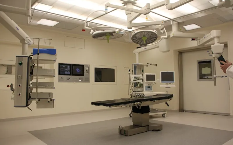
Magnetic Resonance Imaging (MRI) is one of the most sophisticated diagnostic imaging technologies available in modern medicine. Unlike X-rays and CT scans, which use ionizing radiation to create images, MRI uses powerful magnetic fields and radio waves to generate detailed cross-sectional images of the body's internal structures. This radiation-free approach makes MRI particularly valuable for repeated scanning, pediatric imaging, and examining soft tissues where it provides unmatched detail. The technology produces images with extraordinary resolution, allowing physicians to visualize organs, tissues, blood vessels, joints, and even the neural pathways of the brain with remarkable clarity.
The physics behind MRI are fascinating. The human body is approximately 60% water, and each water molecule contains hydrogen atoms. When placed in the strong magnetic field of an MRI scanner, the hydrogen atoms in the body align with the magnetic field. Radio frequency pulses are then sent into the body, momentarily knocking these atoms out of alignment. As the atoms realign with the magnetic field, they emit radio signals that are detected by the scanner's receiver coils. Different tissues (muscle, fat, bone, fluid, tumors) release these signals at different rates, creating contrast between tissue types and allowing the creation of detailed anatomical images.
MRI is particularly superior to other imaging modalities for visualizing soft tissues — brain and spinal cord, joints and cartilage, ligaments and tendons, organs (liver, kidneys, heart), blood vessels, and tumors. It can detect subtle abnormalities that may be invisible on X-ray or CT, making it invaluable for diagnosing conditions ranging from torn ligaments and herniated discs to brain tumors and multiple sclerosis. The ability to create images in any plane (axial, sagittal, coronal) and to use specialized sequences for specific tissue characteristics (diffusion, perfusion, spectroscopy) makes MRI the most versatile imaging tool in medicine.
Types of MRI Technology: 1.5T vs 3T vs Open MRI
MRI scanners come in different field strengths, measured in Tesla (T). The field strength determines the image quality, scan speed, and range of advanced imaging sequences available. Understanding the differences helps you evaluate what technology a hospital offers and whether it meets your diagnostic needs. The three most common types in clinical use are 1.5T (standard), 3T (high-field), and open MRI (typically 0.3-1.0T).
1.5T MRI scanners are the workhorse of diagnostic imaging and remain the most widely deployed MRI systems worldwide. They produce excellent image quality suitable for the vast majority of clinical applications — brain imaging, spine evaluation, joint assessment, abdominal scanning, and general musculoskeletal imaging. 1.5T scans are faster and less expensive than 3T scans, and they produce fewer artifacts (distortions) from metal implants and dental work. For most diagnostic purposes, a well-maintained 1.5T scanner with experienced radiologists produces clinically excellent results.
3T MRI scanners operate at double the field strength of 1.5T systems, producing significantly higher resolution images with greater signal-to-noise ratio. This enhanced detail is particularly valuable for neuroimaging (detecting small brain lesions, micro-bleeds, early demyelination), cardiac MRI (visualizing coronary arteries and myocardial viability), breast MRI (detecting small lesions in dense breast tissue), prostate MRI (multiparametric protocols for cancer detection), and musculoskeletal imaging (visualizing small ligament tears and cartilage defects). Many leading international hospitals have invested in 3T MRI technology, making advanced imaging available abroad at a fraction of Western prices.
Open MRI systems use a lower field strength (0.3-1.0T) but provide a much less confining experience for patients who are claustrophobic or cannot fit in a standard cylindrical MRI bore. Open MRI designs use a C-shaped or open-sided magnet that doesn't enclose the patient. While image quality is lower than 1.5T or 3T systems, open MRI is adequate for many basic diagnostic needs and provides a comfortable alternative for claustrophobic patients. Some hospitals also offer wide-bore 1.5T or 3T scanners that provide a compromise — higher field strength with a wider opening (70cm vs standard 60cm) that is more comfortable for larger patients.
When Do You Need an MRI?
MRI is indicated for a wide range of diagnostic purposes, and understanding when an MRI is necessary versus when other imaging might be more appropriate helps you make informed decisions. Common indications include neurological symptoms (headaches, dizziness, seizures, vision changes, weakness, numbness — brain and spine MRI), joint pain and sports injuries (knee, shoulder, hip, ankle — evaluating ligament tears, cartilage damage, meniscus injuries), back and neck pain (spine MRI for disc herniation, spinal stenosis, nerve compression), cancer staging and monitoring (detecting, characterizing, and monitoring tumors throughout the body), and cardiac evaluation (heart structure, function, valve disease, coronary assessment).
MRI is generally preferred over CT when soft tissue detail is critical, when radiation exposure should be minimized (especially in children and young adults), when multiple follow-up scans are anticipated, and when evaluating the brain, spinal cord, joints, or soft tissue organs. CT is generally preferred for trauma assessment, bone fractures, lung imaging, kidney stones, and acute stroke evaluation (CT is faster). Understanding this distinction can help you discuss imaging choices with your referring physician and the diagnostic team abroad.
Cost Comparison by Country
MRI Scan Cost Comparison 2025
| Country | Single Area MRI | Full-Body MRI | MRI with Contrast |
|---|---|---|---|
| USA | $1,000 - $3,500 | $3,000 - $10,000 | $1,500 - $5,000 |
| UK | $500 - $2,000 | $1,500 - $5,000 | $800 - $3,000 |
| Turkey | $200 - $500 | $500 - $1,500 | $300 - $700 |
| India | $100 - $350 | $300 - $1,000 | $150 - $500 |
| Thailand | $250 - $600 | $600 - $1,800 | $350 - $800 |
| South Korea | $300 - $700 | $800 - $2,500 | $400 - $900 |
| Spain | $350 - $800 | $900 - $2,500 | $500 - $1,000 |
Prices include the scan itself and radiologist interpretation. Contrast MRI uses gadolinium-based contrast agent for enhanced visualization. Full-body MRI covers brain, spine, chest, abdomen, and pelvis.
The cost disparity for MRI scans between countries is striking. A single-area MRI (e.g., brain or knee) costs $1,000-$3,500 in the United States, and this often does not include the radiologist's interpretation fee, which can add $200-$500. In Turkey, the same scan using identical technology costs $200-$500 including expert radiologist interpretation. A full-body MRI screening program that costs $3,000-$10,000 in the US is available for $500-$1,500 in Turkey and $300-$1,000 in India. Even accounting for travel costs, the savings are substantial — particularly for patients needing multiple MRI examinations.
Best Hospitals for MRI Scans Abroad
Medipol Mega University Hospital in Istanbul houses one of Turkey's most advanced diagnostic imaging departments, equipped with the latest 3T MRI scanners from Siemens and GE Healthcare. Their radiology team includes fellowship-trained neuroradiologists, musculoskeletal radiologists, and body imaging specialists who provide expert interpretation comparable to leading Western academic medical centers. The hospital's status as a university hospital means ongoing research and education programs that keep their diagnostic protocols at the cutting edge of medical imaging practice.
Memorial Sisli Hospital operates a comprehensive imaging center with both 1.5T and 3T MRI systems, allowing flexible scheduling and appropriate technology matching for each patient's needs. Their radiologists have extensive experience with international patients and provide detailed English-language reports with comparison to previous imaging when available. The hospital's JCI accreditation ensures rigorous quality control for all imaging procedures, from scanner calibration and maintenance to image acquisition protocols and reporting standards.
In India, major hospital chains including Apollo, Fortis, and Max Healthcare operate advanced radiology departments with 3T MRI capability at remarkably affordable prices. Thailand's Bumrungrad International Hospital has one of Asia's most advanced diagnostic imaging suites. South Korea's Samsung Medical Center and Asan Medical Center are known for cutting-edge imaging research and the latest diagnostic technology. All these institutions provide English-language services and cater extensively to international patients.
Need an MRI scan? Get free quotes from hospitals with the latest 3T MRI technology worldwide.
Get Free MRI Quote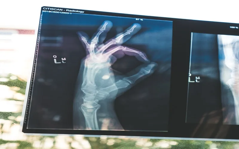
Contrast vs Non-Contrast MRI
MRI scans can be performed with or without contrast agent, and understanding the difference is important for your diagnostic needs. Non-contrast MRI provides excellent soft tissue detail for many applications — brain structural imaging, spine disc evaluation, joint assessment, and general anatomical surveying. Many routine MRI examinations are performed without contrast and provide all the diagnostic information needed.
Contrast-enhanced MRI uses a gadolinium-based contrast agent injected intravenously during the scan. The contrast agent enhances the visibility of blood vessels, inflammation, and abnormal tissue (particularly tumors) by altering the magnetic properties of nearby water molecules. Contrast MRI is particularly important for tumor detection and characterization (distinguishing benign from malignant lesions), evaluating blood vessels (MR angiography), assessing inflammation (joints, spine, brain), post-surgical evaluation (distinguishing scar tissue from recurrent disease), and cardiac MRI (perfusion and viability studies).
Gadolinium contrast agents are generally very safe, with serious adverse reactions occurring in less than 0.01% of administrations. The most common side effects are mild — temporary headache, nausea, or injection site discomfort. However, gadolinium is contraindicated in patients with severe kidney disease (eGFR below 30) due to the risk of nephrogenic systemic fibrosis. Your kidney function will be checked with a blood test before contrast administration. If you have any known allergies or kidney problems, inform the diagnostic team before your appointment.
Bringing Your MRI Results Home
One of the practical considerations when getting an MRI abroad is ensuring your results are useful to your physicians at home. Quality international hospitals make this easy by providing a comprehensive package that includes a detailed radiologist report in English describing all findings, measurements, and diagnostic impressions; DICOM-format digital images on CD, USB drive, or cloud access for viewing and comparison; comparison with any prior imaging you bring; and recommendations for follow-up or additional testing if needed.
To maximize the usefulness of your MRI abroad, bring any previous imaging studies (on CD or in digital format) so the radiologist can compare with baseline images. Share the referral question or clinical concern with the radiologist — knowing what the referring physician is looking for helps focus the interpretation. Request that the report follow the standardized reporting template used in your home country (ACR BI-RADS for breast, PI-RADS for prostate, etc.) for seamless integration with your home healthcare team's workflow.
The quality of an MRI scan depends not just on the machine, but on the expertise of the radiologist interpreting the images. A 1.5T scan read by an expert is far more valuable than a 3T scan read by a generalist. Always ask about the qualifications and specialization of the interpreting radiologist.
American College of Radiology Guidelines
Frequently Asked Questions
How long does an MRI scan take?
A single-area MRI takes 20-45 minutes depending on the body part and whether contrast is used. Full-body MRI takes 60-90 minutes. Cardiac MRI takes 45-60 minutes. You'll need to lie still during the scan. The total appointment time including preparation is usually 60-120 minutes.
Is an MRI scan safe?
MRI is extremely safe as it uses no ionizing radiation. There are no known harmful effects from the magnetic field or radio waves used. However, MRI is contraindicated for patients with certain metallic implants (some pacemakers, cochlear implants, metal fragments). Inform the facility of any implants before scheduling.
Can I get an MRI if I'm claustrophobic?
Yes. Options include: sedation (mild oral or IV sedation), open MRI systems, wide-bore MRI scanners (70cm opening), listening to music during the scan, or having a companion in the room. Discuss your claustrophobia with the hospital beforehand so they can prepare appropriate accommodations.
Will my doctor at home accept MRI results from abroad?
Yes, as long as the images are in standard DICOM format (digital medical imaging standard) and the report is from a qualified radiologist. Most doctors readily accept imaging from accredited international facilities. The key is getting high-quality images and a detailed, well-structured report.
Do I need a doctor's referral for an MRI abroad?
In many countries abroad, you can book an MRI directly without a referral. However, having a referral with a specific clinical question helps the radiologist provide the most targeted interpretation. If you're booking as part of a health checkup package, the examining physician will determine whether MRI is indicated.
What's the difference between a 1.5T and 3T MRI?
3T MRI has double the field strength of 1.5T, producing higher resolution images. 3T is preferred for brain, cardiac, prostate, and breast imaging. 1.5T is excellent for joints, spine, and general imaging. For most diagnostic purposes, both produce clinically adequate images. 3T costs slightly more but is worth it for certain examinations.