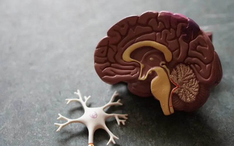
What Is a PET-CT Scan?
Positron Emission Tomography-Computed Tomography (PET-CT) is a revolutionary hybrid imaging technology that combines two powerful diagnostic tools into a single examination. PET imaging detects metabolic activity — how cells in the body are functioning — while CT provides detailed anatomical images showing the exact location and structure of organs and tissues. By fusing these two types of information, PET-CT creates a comprehensive map showing both where abnormal metabolic activity is occurring and precisely where in the body it's located. This dual capability makes PET-CT the most powerful tool available for detecting, staging, and monitoring cancer.
The significance of PET-CT in modern oncology cannot be overstated. Before PET-CT, cancer staging required multiple separate tests — CT scans, bone scans, ultrasounds, and sometimes exploratory surgery — to determine the extent of disease. PET-CT can assess the entire body in a single examination, identifying the primary tumor, lymph node involvement, and distant metastases with remarkable accuracy. Studies have shown that PET-CT changes the treatment plan in 30-40% of cancer patients compared to conventional imaging alone, either by upstaging the disease (finding more extensive cancer than previously known) or downstaging it (finding less extensive disease than feared).
PET-CT technology is widely available at leading hospitals worldwide, with the same scanner models (Siemens Biograph, GE Discovery, Philips Vereos) used in top cancer centers globally. The quality of the scan depends not on geography but on the quality of the equipment, the expertise of the nuclear medicine physicians interpreting the images, and the protocols used for image acquisition. JCI-accredited hospitals abroad operate PET-CT scanners that are identical to those used at Memorial Sloan Kettering, MD Anderson, or the Mayo Clinic — at a fraction of the cost. This democratization of advanced imaging technology through medical tourism gives patients worldwide access to gold-standard cancer diagnostics.
How PET-CT Works: The Science Explained
The PET component of a PET-CT scan works by detecting the metabolic activity of cells. Cancer cells have significantly higher metabolic rates than normal cells — they consume more glucose (sugar) to fuel their rapid growth and division. Before the scan, the patient receives an intravenous injection of a radiotracer, most commonly fluorodeoxyglucose (FDG), which is a glucose molecule labeled with a small amount of radioactive fluorine-18. This radiotracer circulates through the body and is absorbed by cells in proportion to their metabolic activity — cancer cells, with their high glucose appetite, absorb significantly more FDG than normal surrounding tissue.
As the radioactive fluorine-18 in the FDG decays, it emits positrons (positively charged electrons). When a positron encounters an electron in the tissue, both particles are annihilated, producing two gamma photons that travel in exactly opposite directions. The PET scanner's ring of detectors captures these paired photons and uses sophisticated computer algorithms to calculate the exact location of each annihilation event, building up a three-dimensional map of metabolic activity throughout the body. Areas of high FDG uptake appear as bright 'hot spots' on the PET image, potentially indicating cancer, infection, inflammation, or other conditions with elevated metabolic activity.
The CT component provides the anatomical roadmap. While the PET image shows where metabolic hot spots are, the CT image shows exactly what anatomical structures are involved. The fusion of PET and CT data allows physicians to say not just 'there is abnormal metabolic activity' but 'the abnormal activity is located in the right lower lobe of the lung, measuring 2.3cm, with additional activity in the mediastinal lymph nodes' — information that is critical for treatment planning. Modern PET-CT scanners perform both scans simultaneously, with the patient lying on the same table, ensuring perfect anatomical alignment between the two datasets.
When Is PET-CT Recommended?
PET-CT is most commonly used in oncology for cancer detection, staging, treatment response assessment, and surveillance for recurrence. Initial staging is one of the most important applications — when cancer has been diagnosed, PET-CT helps determine the extent of disease throughout the body, which directly influences treatment decisions. For example, a lung cancer that appears localized on CT might be shown by PET-CT to have already spread to distant lymph nodes or organs, changing the treatment approach from surgery to systemic therapy.
Specific cancers where PET-CT is considered essential include lung cancer (staging and treatment response), lymphoma (staging, interim response, end-of-treatment assessment), melanoma (staging of advanced disease), head and neck cancers (staging and radiation planning), esophageal cancer (staging), colorectal cancer (staging of recurrent disease), and breast cancer (staging of locally advanced or metastatic disease). PET-CT is also increasingly used for prostate cancer (using specialized PSMA tracers rather than FDG), cervical cancer, and sarcomas.
Beyond oncology, PET-CT has important applications in cardiology (assessing myocardial viability after heart attack, detecting cardiac sarcoidosis), neurology (evaluating dementia and distinguishing Alzheimer's from other causes, localizing seizure foci for epilepsy surgery), and infectious disease (identifying occult infections and inflammatory conditions like vasculitis). However, oncological applications account for approximately 90% of all PET-CT examinations performed worldwide.
- Cancer Staging: Determining the extent of disease at initial diagnosis — essential for treatment planning
- Treatment Response: Assessing whether chemotherapy or radiation is working by measuring changes in tumor metabolism
- Recurrence Detection: Identifying cancer recurrence earlier than conventional imaging — metabolic changes precede structural changes
- Radiation Planning: Defining tumor boundaries for precise radiation therapy targeting
- Unknown Primary: Identifying the primary tumor when cancer has been found in lymph nodes or metastatic sites but the origin is unknown
- Characterizing Lesions: Distinguishing benign from malignant lesions — a nodule that is metabolically active is more likely malignant
Cost Comparison by Country
PET-CT Scan Cost Comparison 2025
| Country | Whole-Body PET-CT | PET-CT + Consultation | Savings vs USA |
|---|---|---|---|
| USA | $3,000 - $12,000 | $4,000 - $15,000 | — |
| UK | $1,500 - $5,000 | $2,000 - $6,000 | Up to 60% |
| Turkey | $500 - $1,200 | $700 - $1,500 | Up to 90% |
| India | $300 - $800 | $400 - $1,000 | Up to 92% |
| Thailand | $600 - $1,500 | $800 - $2,000 | Up to 85% |
| South Korea | $800 - $2,000 | $1,000 - $2,500 | Up to 80% |
| Spain | $1,000 - $2,500 | $1,200 - $3,000 | Up to 75% |
Prices include the radiotracer (FDG), the scan, and radiologist/nuclear medicine physician interpretation. Some hospitals include oncologist consultation in their package price.
PET-CT is one of the most expensive diagnostic tests in medicine, primarily because of the cost of the radiotracer (FDG must be produced fresh at a cyclotron facility, as fluorine-18 has a short half-life of 110 minutes), the sophisticated and expensive scanner equipment, and the specialized nuclear medicine physician expertise required for interpretation. In the United States, a whole-body PET-CT scan costs $3,000-$12,000, with prices varying significantly by region and facility. Insurance coverage for PET-CT requires meeting specific clinical indications and prior authorization.
The savings available abroad are among the most dramatic in all of medical tourism. Turkey and India offer PET-CT scans at 85-92% less than US prices, using the same scanner technology and internationally trained nuclear medicine physicians. A PET-CT that costs $6,000 at a US cancer center can be performed for $500-$1,200 in Turkey or $300-$800 in India — including expert interpretation and often an oncologist consultation. For cancer patients who need multiple PET-CT scans during treatment (staging, interim assessment, post-treatment evaluation), the cumulative savings of getting these scans abroad can be enormous.
Preparation & What to Expect
Preparing for a PET-CT scan requires specific attention to diet and activity in the hours before the examination. You must fast for 4-6 hours before the scan — no food, sweetened drinks, or chewing gum (even sugar-free gum can trigger insulin release). Water is allowed and encouraged. Avoid strenuous exercise for 24 hours before the scan, as exercise increases glucose uptake in muscles, potentially creating false positive results. If you have diabetes, special preparation instructions will be provided — your blood sugar must be within acceptable range (typically below 200 mg/dL) for the scan to be accurate.
On the day of the scan, you'll arrive at the nuclear medicine department and have your blood glucose checked. The FDG radiotracer is then injected intravenously, and you'll rest quietly for 45-60 minutes in a warm, dimly lit room while the tracer distributes throughout your body. Remaining still and avoiding talking is important during this uptake period to minimize FDG absorption by muscles. After the uptake period, you'll be positioned on the scanner table and the PET-CT scan itself takes approximately 20-35 minutes, during which you need to lie still while the scanner moves along your body.
After the scan, you can resume normal activities immediately. The radioactive tracer is naturally eliminated from your body through urination within 6-12 hours. Drinking plenty of water after the scan helps flush the tracer more quickly. The radiation exposure from a standard PET-CT is approximately 7-25 mSv, comparable to a conventional CT scan and considered safe for diagnostic purposes. You may be asked to avoid close contact with pregnant women and small children for a few hours after the scan as a precaution, although the radiation levels are very low.
Best Hospitals for PET-CT Abroad
Acıbadem Maslak Hospital in Istanbul operates one of Turkey's most advanced nuclear medicine departments, equipped with the latest PET-CT technology including Siemens Biograph Vision and GE Discovery MI systems. Their nuclear medicine team includes fellowship-trained physicians with extensive experience in oncological PET-CT interpretation, cardiac PET, and neurological imaging. The hospital's multidisciplinary tumor board integrates PET-CT findings into comprehensive cancer care plans, making it an excellent choice for patients seeking both diagnosis and treatment.
Liv Hospital Istanbul offers state-of-the-art PET-CT services within its comprehensive oncology center. The hospital uses the latest digital PET-CT technology, which provides higher resolution images with lower radiation dose compared to older analog systems. Their nuclear medicine department works closely with the oncology, radiation therapy, and surgical teams to ensure PET-CT findings are integrated into treatment planning. For international patients, Liv Hospital provides seamless coordination between PET-CT scheduling, oncology consultation, and results reporting.
Need a PET-CT scan for cancer diagnosis or staging? Get free quotes from hospitals with the latest PET-CT technology.
Get Free PET-CT Quote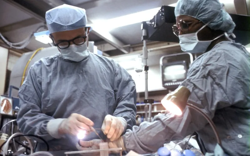
Interpreting PET-CT Results
PET-CT results are expressed using the Standardized Uptake Value (SUV), which quantifies how much FDG a particular area of the body has absorbed relative to the overall body distribution. The maximum SUV (SUVmax) of a lesion is the most commonly used metric. Generally, an SUVmax below 2.5 suggests a benign process, while values above 2.5 raise suspicion for malignancy — though this threshold is not absolute and varies by tissue type. Some benign conditions (infection, inflammation, granulomatous disease) can produce high SUV values, while some cancers (particularly slow-growing tumors like well-differentiated thyroid cancer or low-grade lymphoma) may show low SUV values.
The PET-CT report from a quality hospital abroad will include detailed description of all areas of abnormal FDG uptake with SUVmax measurements, anatomical localization using the fused PET-CT images, comparison with previous PET-CT or other imaging studies if available, assessment of treatment response using standardized criteria (such as Deauville criteria for lymphoma or PERCIST criteria for solid tumors), overall impression with differential diagnosis, and recommendations for further evaluation or follow-up. This comprehensive reporting format is compatible with treatment protocols at any cancer center worldwide.
It's important to understand that PET-CT is a powerful but imperfect tool. False positives (areas that light up on PET but are not cancer) can occur due to infection, inflammation, granulomatous disease, brown fat activation, muscle tension, and recent surgery. False negatives (cancer that doesn't show on PET) can occur with slow-growing tumors, small lesions below the resolution limit (typically less than 7-8mm), brain metastases (the brain normally has high glucose metabolism, masking tumor uptake), and mucinous tumors. These limitations should be discussed with your oncologist when interpreting results.
PET-CT has revolutionized cancer care by allowing us to see cancer's metabolic fingerprint throughout the body in a single examination. It has fundamentally changed how we stage cancer, plan treatment, and monitor response — improving outcomes for millions of patients worldwide.
Journal of Nuclear Medicine, Cancer Imaging Guidelines
Frequently Asked Questions
Is a PET-CT scan safe?
Yes, PET-CT is safe for diagnostic purposes. The radiation exposure (7-25 mSv) is comparable to a standard CT scan and well within accepted safety limits. The radiotracer (FDG) is naturally eliminated from your body within 6-12 hours. Allergic reactions to FDG are extremely rare. PET-CT should be avoided during pregnancy.
How accurate is PET-CT for detecting cancer?
PET-CT has a sensitivity of 85-95% and specificity of 80-90% for most cancer types, making it the most accurate whole-body cancer imaging modality available. It's particularly accurate for lung cancer, lymphoma, and melanoma staging. However, it has limitations for small lesions (<8mm), brain metastases, and slow-growing tumors.
Can PET-CT detect all types of cancer?
PET-CT detects most cancer types with high accuracy, but some cancers have lower FDG avidity (glucose uptake). Cancers well-detected include lung, lymphoma, melanoma, head and neck, esophageal, and colorectal. Less well-detected include prostate (use PSMA-PET instead), renal cell carcinoma, low-grade lymphoma, and well-differentiated thyroid cancer. Your oncologist will advise the most appropriate tracer.
How long does a PET-CT scan take?
The total appointment takes approximately 2-3 hours. This includes: blood glucose check (5 minutes), FDG injection (5 minutes), uptake rest period (45-60 minutes), the actual scan (20-35 minutes), and a short observation period. The uptake rest period, where you relax while the tracer distributes, is the longest part.
Can I eat before a PET-CT scan?
No, you must fast for 4-6 hours before a PET-CT scan. Eating raises blood sugar and insulin levels, which interfere with FDG uptake by cancer cells and can produce inaccurate results. Water is allowed and encouraged. Avoid strenuous exercise for 24 hours before the scan.
Will my insurance cover a PET-CT scan abroad?
Most US insurance plans do not cover PET-CT scans performed abroad. However, the cost of a PET-CT scan in Turkey or India ($300-$1,200) is often less than the insurance copay/deductible for the same scan in the US. Some international insurance plans and global health insurance policies do cover imaging abroad at accredited facilities.